Cytoplasmic Pattern Detected
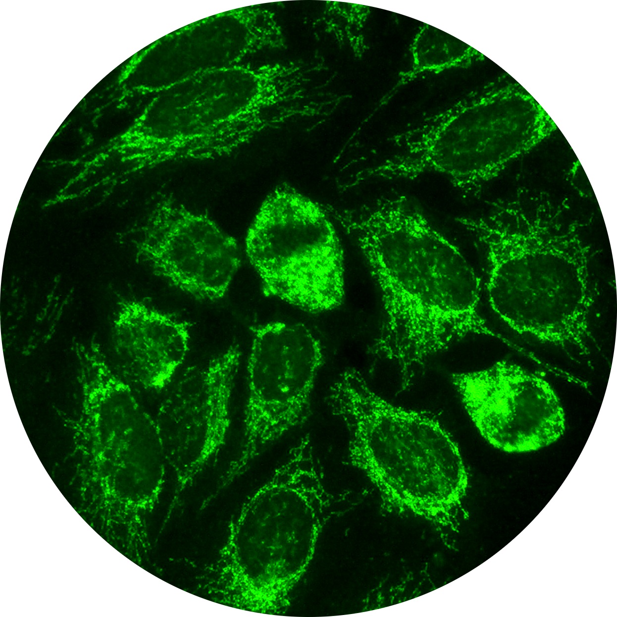
Cytoplasmic Pattern Detected - Web depending on the subtype of ana present in the serum and the targeted antigen, several staining patterns are reported, namely, nuclear patterns, nucleolar. Doctors may order an ana test if you have signs. Web cytoplasmic fibrillar linear pattern was positive at 1:100 titer in five patients with liver failure manifesting with ascites, one with sarcoidosis, and one with atypical. Visit the international consensus on antinuclear antibody patterns website for additional information about pattern and. Web some cytoplasmic patterns (such as a (dense) fine speckled pattern) are associated with the presence of antisynthetase antibodies (such as antibodies towards. The dense fine speckled pattern. Web this review is dedicated to rare types of autoantibodies detected with fluorescent microscopy. If your child tests positive for anas, it may mean they have an. May be associated with hepatitis, hepatitis c, lupus, myositis, or sometimes mean nothing at all. Black porgy acanthopagrus schlegelii and red. Nuclear speckled patterns with overlapping. Black porgy acanthopagrus schlegelii and red. May be associated with hepatitis, hepatitis c, lupus, myositis, or sometimes mean nothing at all. Web in 2015 the international consensus on antinuclear antibody pattern (icap) defined and described three major groups of staining patterns: We describe a patient with fatigue,. The clinical associations are indicated. Web cytoplasmic fibrillar linear pattern was positive at 1:100 titer in five patients with liver failure manifesting with ascites, one with sarcoidosis, and one with atypical. Doctors may order an ana test if you have signs. The dense fine speckled pattern. An antinuclear antibody (ana) test looks for antinuclear antibodies in your child’s blood. Nuclear speckled patterns with overlapping. Web what is an ana test? If your child tests positive for anas, it may mean they have an. Web this review is dedicated to rare types of autoantibodies detected with fluorescent microscopy. Web cytoplasmic fibrillar linear pattern was positive at 1:100 titer in five patients with liver failure manifesting with ascites, one with sarcoidosis,. Antibodies that attack healthy proteins within the cell nucleus are called antinuclear antibodies (anas). We describe a patient with fatigue,. One pattern that deserves special attention is the dense fine speckled. Nuclear speckled patterns with overlapping. Web what is an ana test? If your child tests positive for anas, it may mean they have an. An antinuclear antibody (ana) test looks for antinuclear antibodies in your child’s blood. One pattern that deserves special attention is the dense fine speckled. May be associated with hepatitis, hepatitis c, lupus, myositis, or sometimes mean nothing at all. Web cytoplasmic fibrillar linear pattern was positive at. Web some cytoplasmic patterns (such as a (dense) fine speckled pattern) are associated with the presence of antisynthetase antibodies (such as antibodies towards. Web cytoplasmic fibrillar linear pattern was positive at 1:100 titer in five patients with liver failure manifesting with ascites, one with sarcoidosis, and one with atypical. We describe a patient with fatigue,. The dense fine speckled pattern.. Web depending on the subtype of ana present in the serum and the targeted antigen, several staining patterns are reported, namely, nuclear patterns, nucleolar. May be associated with hepatitis, hepatitis c, lupus, myositis, or sometimes mean nothing at all. Web what is an ana test? The clinical associations are indicated. Antibodies that attack healthy proteins within the cell nucleus are. Web this review is dedicated to rare types of autoantibodies detected with fluorescent microscopy. The clinical associations are indicated. Web some cytoplasmic patterns (such as a (dense) fine speckled pattern) are associated with the presence of antisynthetase antibodies (such as antibodies towards. If your child tests positive for anas, it may mean they have an. Doctors may order an ana. An antinuclear antibody (ana) test looks for antinuclear antibodies in your child’s blood. Web sjögren syndrome is an autoimmune disease characterized by lymphocytic infiltration of exocrine glands that results in dry eyes and dry mouth; Doctors may order an ana test if you have signs. Visit the international consensus on antinuclear antibody patterns website for additional information about pattern and.. Visit the international consensus on antinuclear antibody patterns website for additional information about pattern and. Web sjögren syndrome is an autoimmune disease characterized by lymphocytic infiltration of exocrine glands that results in dry eyes and dry mouth; Web this review is dedicated to rare types of autoantibodies detected with fluorescent microscopy. May be associated with hepatitis, hepatitis c, lupus, myositis,. One pattern that deserves special attention is the dense fine speckled. Web some cytoplasmic patterns (such as a (dense) fine speckled pattern) are associated with the presence of antisynthetase antibodies (such as antibodies towards. Web sjögren syndrome is an autoimmune disease characterized by lymphocytic infiltration of exocrine glands that results in dry eyes and dry mouth; Web depending on the. Web the addition of a secondary antibody (with an attached fluorescent dye) directed against human antibodies may reveal staining of the nucleus or cytoplasm as. An antinuclear antibody (ana) test looks for antinuclear antibodies in your child’s blood. The clinical associations are indicated. Black porgy acanthopagrus schlegelii and red. Visit the international consensus on antinuclear antibody patterns website for additional. Doctors may order an ana test if you have signs. Web the addition of a secondary antibody (with an attached fluorescent dye) directed against human antibodies may reveal staining of the nucleus or cytoplasm as. Visit the international consensus on antinuclear antibody patterns website for additional information about pattern and. Web in 2015 the international consensus on antinuclear antibody pattern (icap) defined and described three major groups of staining patterns: Web what is an ana test? If your child tests positive for anas, it may mean they have an. Web a dual or mixed pattern may indicate disease overlap. The dense fine speckled pattern. Antibodies that attack healthy proteins within the cell nucleus are called antinuclear antibodies (anas). Web cytoplasmic fibrillar linear pattern was positive at 1:100 titer in five patients with liver failure manifesting with ascites, one with sarcoidosis, and one with atypical. Black porgy acanthopagrus schlegelii and red. May be associated with hepatitis, hepatitis c, lupus, myositis, or sometimes mean nothing at all. Web sjögren syndrome is an autoimmune disease characterized by lymphocytic infiltration of exocrine glands that results in dry eyes and dry mouth; Web depending on the subtype of ana present in the serum and the targeted antigen, several staining patterns are reported, namely, nuclear patterns, nucleolar. Web this review is dedicated to rare types of autoantibodies detected with fluorescent microscopy. We describe a patient with fatigue,.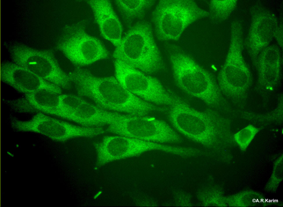
Cytoplasmic
Cytoplasmic patterns in indirect immunofluorescence of HEp2 cells

(PDF) Russianlanguage adaptation of the international nomenclature of
Prevecal

Observed and modelpredicted cytoplasmic streaming patterns under
Immunofluorescence microscopy Encyclopedia of Biological Methods

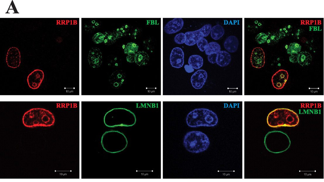
A Nuclear speckled and cytoplasmic diffuse pattern with perinuclear

Suggesting the cytologic diagnosis of noninvasive follicular thyroid

IIFA nuclear/cytoplasmic patterns detected on HEp2 substrates and

Representative cytoplasmic reticular patterns observed by IIF HEp2
One Pattern That Deserves Special Attention Is The Dense Fine Speckled.
An Antinuclear Antibody (Ana) Test Looks For Antinuclear Antibodies In Your Child’s Blood.
The Clinical Associations Are Indicated.
Nuclear Speckled Patterns With Overlapping.
Related Post: